1944 Smithsonian Report Of
The Rife Universal Microscope
|
THE SMITHSONIAN REPORT
From the Annual Report of the Board of Regents of The Smithsonian Institution
Vol. 237 FEBRUARY, 1944 NO.2
THE NEW MICROSCOPES.
A Discussion by 1944R. E. SEIDEL, M.D. AND M. ELIZABET WINTER, Philadelphia, Pa.
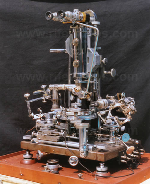
The Universal Microscope
It is only a reasonable supposition, but already, in one instance, a very successful and highly commendable achievement on the part of Dr. Royal Raymond Rife of San Diego, California, who, for many years, has built and worked with light microscopes which far surpass the theoretical limitations of the ordinary variety of instrument, all the Rife scopes possessing superior ability to attain high magnification with accompanying high resolution.
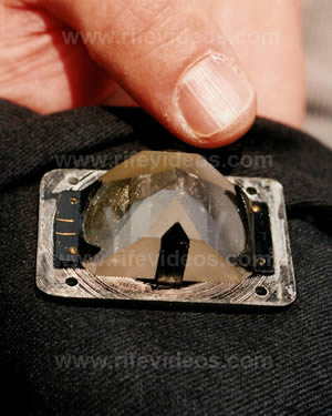
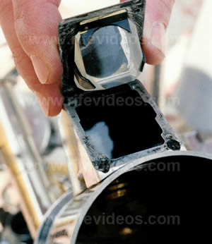
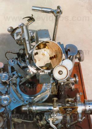
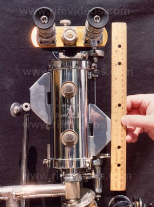
The largest and most powerful of these, the Universal Microscope, developed in 1933, consists of 5,682 parts and is so called because of its adaptability in all fields of microscopical work, being fully equipped with separate substage condenser units for transmitted and monochromatic beam dark-field, polarized, and slit-ultra illumination, including also a special device for crystallography. The entire optical system of lenses and prisms as well as the illuminating units are made of block-crystal quartz, quartz being especially transparent to ultraviolet radiations.
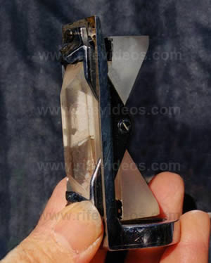
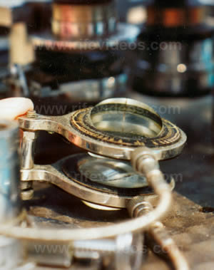
This illuminating unit used for examining the filterable forms of disease organisms contains 14 lenses and prisms, 3 of which are in the high-intensity incandescent lamp, 4 in the Risley prism, and 7 in the achromatic condenser which, incidentally, has a numerical aperture of 1.40. Between the source of light and the specimen are subtended two circular, wedge-shaped, block-crystal quartz prisms for the purpose of polarizing the light passing through the specimen, polarization being the practical application of the theory that light waves vibrate in all planes perpendicular to the direction in which they are propagated.
Therefore, when light comes into contact with a polarizing prism, it is divided or split into two beams, one of which is refracted to such an extent that it is reflected to the side of the prism without, of course, passing through the prism while the second ray, bent considerably less, is thus enabled to pass through the prism to illuminate the specimen.
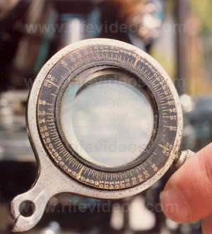
When the quartz prisms on the universal microscope, which may be rotated with vernier control through 360 degrees, are rotated in opposite directions, they serve to bend the transmitted beams of light at variable angles of incidence while, at the same time, a spectrum is projected up into the axis of the microscope, or rather a small portion of the spectrum to the other, going all the way from the infrared to the ultraviolet.
Now, when that portion of the spectrum is reached in which both the organism and the color band vibrate in exact accord, one with the other, a definite characteristic spectrum is emitted by the organism.
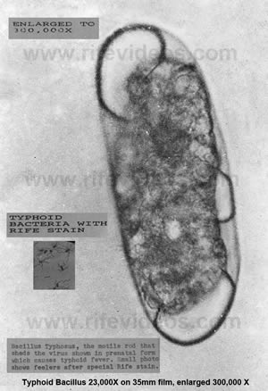
In the case of the filter-passing form of the Bacillus Typhosus, for instance, a blue spectrum is emitted and the plane of polarization deviated plus (+) 4.8 degrees.
The predominating chemical constituents of the organism are next ascertained after which the quartz prisms are adjusted or set, by means of vernier control, to minus (-) 4.8 degrees (again in the case of the filter-passing form of the Bacillus Typhosus) so that the opposite angle of refraction may be obtained.
A monochromatic beam of light, corresponding exactly to the frequency of the organism (for Dr. Rife has found that each disease organism responds to and has a definite wave length, a fact confirmed by British medical research workers) is then sent up through the specimen and the direct transmitted light, thus enabling the observer to view the organism stained in its true chemical color and revealing its own individual structure in a field which is brilliant with light.
The objectives used on the universal microscope are a 1.12 dry lens, a 1.16 water immersion, a 1.18 oil immersion, and a 1.25 oil immersion. The rays of light refracted by the specimen enter the objective and are then carried up the tube in parallel rays through 21 light bends to the ocular, a tolerance of less than one wave length of visible light only being permitted in the core beam, or chief ray, of illumination.
Now, instead of the light rays starting up the tube in a parallel fashion, tending to converge as they rise higher and finally crossing each other, arriving at the ocular separated by considerable distance as would be the case with an ordinary microscope, in the universal tube the rays also start their rise parallel to each other but, just as they are about to pull them out parallel again, another prism being inserted each time the rays are about ready to cross.
These prisms, inserted in the tube, which are adjusted and held in alignment by micrometer screws of 100 threads to the inch in special tracks made of magnelium (magnelium having the closest coefficient of expansion of any metal to quartz), are separated by a distance of only 30 millimeters.
Thus, the greatest distance that the image in the universal microscope is projected through any one media, either quartz or air, is 30 millimeters instead of the 160, 180, or 190 millimeters as in the empty or air-filled tubes of an ordinary microscope, the total distance which the light rays travel zigzag fashion through the universal tube being 449 millimeters, although the physical length of the tube itself is 229 millimeters.
It will be recalled that if one pierces a black strip of paper or cardboard with the point of a needle and then brings the card up close to the eye so that the hole is in the optic axis, a small brilliantly lighted object will appear larger and clearer, revealing more fine detail, than if it were viewed from the same distance without the assistance of the card.
This is explained by the fact that the beam of light passing through the card is very narrow, the rays entering the eye, therefore, being practically parallel, whereas without the card the beam of light is much wider and the diffusion circles much larger. It is this principle of parallel rays in the universal microscope and the resultant shortening of projection distance between any two blocks or prisms plus the fact that objectives can thus be substituted for oculars, these "oculars" being three matched pairs of 10 millimeter, 7 millimeter, and 4 millimeter objectives in short mounts, which would make possible not only the unusually high magnification and resolution but which serve to eliminate all distortion as well as all chromatic and spherical aberration.
Quartz slides with especially thin quartz cover glasses are used when a tissue section or culture slant is examined, the tissue section itself also being very thin. An additional observational tube and ocular which yield a magnification of 1,800 diameters are provided so that that portion of the specimen which is desired to be examined may be located so that the observer can adjust himself more readily when viewing a section at a high magnification.
The universal stage is a double rotating stage graduated through 360 degrees in quarter-minute arc divisions, the upper segment carrying the mechanical stage having a movement of 40 degrees, plus or minus. Heavily constructed joints and screw adjustments maintain rigidity of the microscope which weighs 200 pounds and stands 24 inches high, the bases of the scope being nickel cast-steel plates, accurately surfaced, and equipped with three leveling screws and two spirit levels set at angles of 90 degrees. The coarse adjustment, a block thread screw with 40 threads to the inch, slides in a 1 1/2 dovetail which gibes directly onto the pillar post. The weight of the quadruple nosepiece and the objective system is taken care of by the intermediate adjustment at the top of the body tube. The stage, in conjunction with a hydraulic lift, acts as a lever in operating the fine adjustment. A 6-gauge screw having 100 threads to the inch is worked through a gland into a hollow, glycerine-filled post, the glycerine being displaced and replaced at will as the screw is turned clockwise or anticlockwise, allowing a 5-to-1 ratio on the lead screw. This, accordingly, assures complete absence of drag and inertia. The fine adjustment being 700 times more sensitive then that of ordinary microscopes, the length of time required to focus the universal ranges up to 1 1/2 hours which, while on first consideration, may seem a disadvantage, is after all but a slight inconvenience when compared with the many years of research and the hundreds of thousands of dollars spent and being spent in an effort to isolate and to look upon disease-causing organisms in their true form.
Working together back in 1931 and using one of the smaller Rife microscope having a magnification and resolution of 17,000 diameters, Dr. Rife and Dr. Arthur Isaac Kendall, of the department of bacteriology of Northwestern University Medical School, were able to observe and demonstrate the presence of the filter-passing forms of Bacillus Typhosus. An agar slant culture of the Rawlings strain of Bacillus Typhosus was first prepared by Dr. Kendall and inoculated into 6 cc of "Kendall" K Medium, a medium rich in protein but poor in peptone and consisting of 100 mg. of dried hog intestine and 6 cc of tyrode solution (containing neither glucose nor glycerine) which mixture is shaken well so as to moisten the dried intestine powder and then sterilized in the autoclave, 15 pounds for 15 minutes, alterations of the medium being frequently necessary depending upon the requirements for different organisms. Now, after a period of 18 hours in this K Medium, the culture was passed through a Berkefeld "N" filter, a drop of the filtrate being added to another 6 cc. of K Medium and incubated at 37 degrees C. Forty-eight hours later this same process was repeated, the "N" filter again being used, after which it was noted that the culture no longer responded to peptone medium, growing now only in the protein medium. When again, within 24 hours, the culture was passed through a filter-the finest Berkefeld "W" filter, a drop of the filtrate was once more added to 6 cc.of K Medium and incubated at 37 degrees c., a period of 3 days elapsing before a new culture was transferred to K Medium and yet another 3 days before a new culture was prepared. Then, viewed under an ordinary microscope, these cultures were observed to be turbid and to reveal no bacilli whatsoever. When viewed by means of dark-field illumination and oil-immersion lens, however, the presence of small, actively motile granules was established, although nothing at all of their individual structure could be ascertained. Another period of 4 days was allowed to elapse before these cultures were transferred to K Medium and incubated at 37 degrees C for 24 hours when they were then examined under the Rife microscope where, as was mentioned earlier, the filterable typhoid bacilli, emitting a blue spectrum, caused the plane of polarization to be deviated plus 4.8 degrees. Then when the opposite angle of refraction was obtained by means of adjusting the polarizing prisms to minus 4.8 degrees and the cultures illuminated by a monochromatic beam coordinated in frequency with the chemical constituents of the typhoid bacillus, small oval actively motile, bright turquoise-blue bodies were observed at a magnificatinn of 5,000 diameters, in high contrast to the colorless and motionless debris of the medium. These observations were repeated eight times, the complete absence of these bodies in uninoculated control K Media also being noted.
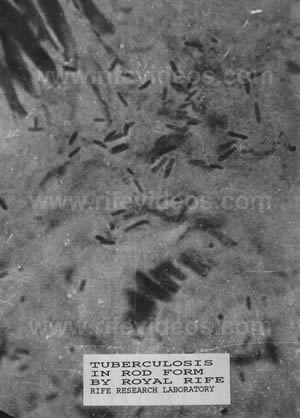
To further confirm their findings, Drs. Rife and Kendall next examined 18-hour-old cultures which had been inoculated into K Medium and incubated at 37 degrees C., since it is just at this stage of growth in this medium and at this temperature that the cultures become filterable. And, just as had been anticipated, ordinary dark-field examination revealed unchanged, long, actively motile bacilli; bacilli having granules within their substance; and free-swimming, actively motile granules; while under the Rife microscope were demonstrated the same long, unchanged, almost colorless bacilli; bacilli, practically colorless, inside and at one end of which was a turquoise-blue granule resembling the filterable forms of the typhoid bacillus; and free-swimming, small, oval, actively motile,turquoise-blue granules. By transplanting the cultures of the filter-passing organisms or virus into a broth, they were seen to change over again into their original rodlike forms.
At the same time that these findings of Drs. Rife and Kendall were confirmed by Dr. Edward C. Rosenow, of the Mayo Foundation, the magnification with accompanying resolution of 8,000 diameters of the Rife microscope, operated by Dr. Rife, was checked against a dark-field oil-immersion scope operated by Dr. Kendall and an ordinary 2-mm. oil-immersion objective, x 10 ocular, Zeiss scope operated by Dr.Rosenow at a magnification of 900 diameters. Examinations of gram and safranin-stained films of culture of Bacillus typhosus, gram and safranin-stained films of blood and of the sediment of the spinal fluid from a case of acute poliomyelitis were made with the result that bacilli, streptococci, erythrocytes, polymorphonuclear leukocytes, and lymphocytes measuring nine times the diameter of the same specimens observed under the Zeiss scope at a magnification and resolution of 900 diameters, were revealed with unusual clarity. Seen under the dark-field microscope were moving bodies presumed to be the filterable turquois-blue bodies of the typhoid bacillus which, as Dr.Rosenow has declared in his report (Observations on filter-passing forms of Eberthella-typhi-Bacillus typhosus - and of the streptococcus from poliomyelitis, Proc.Staff Meeting Mayo Clinic, July 13, 1932), were so "unmistakably demonstrated" with Rife microscope, while under the Zeiss scope stained and hanging-drop preparations of clouded filtrate culture were found to be uniformly negative. With the Rife microscope also were demonstrated brownish-gray cocci and diplococci in hanging-drop preparations of the filtrates of streptococcus from poliomyelitis. These cocci and diplococci, similar in size and shape to those seen in the culture although of more uniform intensity, and characteristic of the medium in which they had been cultivated, were surrounded by a clear halo about twice the width of that at the margins of the debris and of the Bacillus typhosus. Stained films of filtrates and filtrate sediments examined under the Zeiss microscope, and hanging-drop, dark-field preparations revealed no organisms, however. Brownish-gray cocci and diplococci of the exact same size and density as those observed in the filtrates of the streptococcus cultures were also revealed in hanging-drop preparations of the virus of poliomyelitis underthe Rife microscope, while no organisms at all could be seen in either the stained films of filtrates and filtrate sediments examined with the Zeiss scope or in hanging-drop preparations examined by means of the dark-field. Again using the Rife microscope at a magnification of 8,000 diameters, numerous nonmotile cocci and diplococci of a bright-to-pale pink in color were seen in hanging-drop preparations of filtrates of Herpes encephalitic virus. Although these were observed to be comparatively smaller then the cocci and diplococci of the streptococcus and poliomyelitis viruses, they were shown to be of fairly even density, size and form and surrounded by a halo. Again, both the dark-field and Zeiss scopes failed to reveal any organisms, and none of the three microscopes disclosed the presence of such diplococci in hanging-drop preparation of the filtrate of a normal rabbit brain. Dr. Rosenow has since revealed these organisms with the ordinary microscope at a magnification of 1,000 diameters by means of his special staining method and with the electron microscope at a magnification of 12,000 diameters. Dr. Rosenow has expressed the opinion that the inability to see these and other similarly revealed organisms is due, not necessarily to the minuteness of the organisms, but rather to the fact that they are of a nonstaining, hyaline structure. Results with the Rife microscopes, he thinks, are due to the "ingenious methods employed rather than to excessively high magnification." He has declared also, in the report mentioned previously, that "Examination under the Rife microscope of specimens containing objects visible with the ordinary microscope, leaves no doubt of the accurate visualization of objects or particulate matter by direct observation at the extremely high magnification obtained with this instrument."
Exceedingly high powers of magnification with accompanying high powers of resolution may be realized with all of the Rife microscopes, one of which, having magnification and resolution up to 18,000 diameters, is now being used at the British School of Tropical Medicine in England. In a recent demonstration of another of the smaller Rife scopes (May 16, 1942) before a group of doctors including Dr. J.H. Renner, of Santa Barbara, Calif.; Dr. Roger A. Schmidt, of San Francisco, Calif.; Dr. Lois Bronson Slade, of Alameda, Calif.; Dr. Lucile B. Larkin, of Bellingham, Wash.; Dr. E. F. Larkin, of Bellingham, Wash.; and Dr. W. J. Gier, of San Diego, Calif., a Zeiss ruled grading was examined, first under an ordinary commercial microscope equipped with a 1.8 high dry lens and X 10 ocular, and then under the Rife microscope. Whereas 50 lines were revealed with the commercial instrument and considerable aberration, both chromatic and spherical noted, only 5 lines were seen with the Rife scope, these 5 lines being so highly magnified that they occupied the entire field, without any aberration whatsoever being apparent. Dr. Renner, in a discussion of his observations, stated that "The entire field to its very edges and across the center had a uniform clearness that was not true on the conventional instrument." Following the examination of the grading, an ordinary unstained blood film was observed under the same two microscopes. In this instance, 100 cells were seen to spread throughout the field of the commercial instrument while but 10 cells filled the field of the Rife scope.
The universal microscope, of course, is the most powerful Rife scope, possessing a resolution of 31,000 diameters and magnification of 60,000 diameters. With this it is possible to view the interior of the 'pin-point' cells, those cells situated between the normal tissue cells and just visible under the ordinary microscope, and to observe the smaller cells which compose the interior of these pin-point cells. When one of these smaller cells in magnified, still smaller cells are seen within its structure. And when one of the still smaller cells, in its turn, is magnified, it, too, is seen to be composed of smaller cells. Each of the 16 times this process of magnification and resolution can be repeated, it is demonstrated that there are smaller cells within the smaller cells, a fact which amply testifies as to the magnification and resolving power obtainable with the universal microscope.
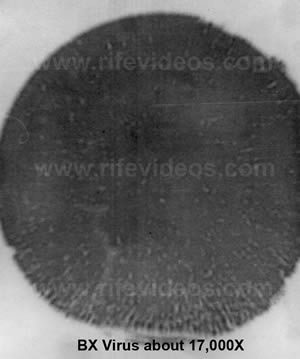
More then 20,000 laboratory cultures of carcinoma were grown and studied over a period of 7 years by Dr. Rife and his assistants in what, at the time, appeared to be a fruitless effort to isolate the filter-passing form, or virus, which Dr. Rife believed to be present in this condition. Then, in 1932, the reactions in growth of bacterial cultures to light from the rare gasses was observed, indicating a new approach to the problem. Accordingly, blocks of tissue one-half centimeter square, taken from an unulcerated breast carcinoma, were placed in a circular glass loop filled with argon gas to a pressure of 14 millimeters, and a current of 5,000 volts applied for 24 hours, after which the tubes were placed in a 2-inch water vacuum and incubated at 37 degrees C. for 24 hours. Using a specially designed 1.12 dry lens, equal in amplitude of magnification to the 2-mm. apochromatic oil-immersion lens, the cultures were then examined under the universal microscope, at a magnification of 10,000 diameters, where very much animated, purplish-red, filterable forms, measuring less then one-twentieth of a micron in dimension, were observed. Carried through 14 transplants from K Medium to K Medium, this B. X. virus remained constant; inoculated into 426 Albino rats, tumors "with all the true pathology of neoplastic tissue" were developed. Experiments conducted in the Rife Laboratories have established the fact that these characteristic diplococci are found in the blood monocytes in 92 percent of all cases of neoplastic diseases. It has also been demonstrated that the virus of cancer, like the viruses of other diseases, can be easily changed from one form to another by means of altering the media upon which it is grown. With the first change in media, the B. X. virus becomes considerably enlarged although its purplish-red color remains unchanged.
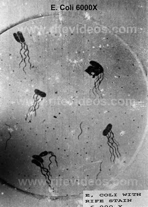
Observation of the organism with an ordinary microscope is made possible by a second alteration of the media. A third change is undergone upon asparagus base media where the B. X. virus is transformed from its filterable state into cryptomyces pleomorphia fungi, these fungi being identical morphologically both microscopically to that of the orchid and of the mushroom. And yet a fourth change may be said to take place when this cryptomyces pleomorphia, permitted to stand as a stock culture for the period of metastasis, becomes the well-known mahogany-colored Bacillus coli.
It is Dr. Rife's belief that all micro-organisms fall into 1 of not more than 10 individual groups (Dr. Rosenow has stated that some of the viruses belong to the group of the streptococcus), and that any alteration of artificial media of slight metabolic variation in tissues will induce an organism of one group to change over into any other organism included in that same group, it being possible, incidentally, to carry such changes in media or tissues to the point where the organisms fail to respond to standard laboratory methods of diagnosis. These changes can be made to take place in as short a period of time as 48 hours. For instance, by altering the media - 4 parts per million per volume - the pure culture of mahogany-colored Bacillus coli becomes the turquoise-blue Bacillus typhosus. Viruses of primordial cells of organisms which would ordinarily require an 8-week incubation period to attain their filterable state, have been shown to produce disease within 3 days' time, proving Dr. Rife's contention that the incubation period of a micro-organism is really only a cycle of reversion.
He states:
"In reality, it is not the bacteria themselves that produce the disease, but we believe it is the chemical constituents of these micro-organisms enacting upon the unbalanced cell metabolism of the human body that in actuality produce the disease. We also believe if the metabolism of the human body is perfectly balanced or poised, it is susceptible to no disease."
In other words, the human body itself is chemical in nature, being comprised of many chemical elements which provide the media upon which the wealth of bacteria normally present in the human system feed. These bacteria are able to reproduce. They, too, are composed of chemicals. Therefore, if the media upon which they feed, in this instance the chemicals or some portion of the chemicals of the human body, become changed from the normal, it stands to reason that these same bacteria, or at least certain numbers of them, will also undergo a change chemically since they are now feeding upon media which are not normal to them, perhaps being supplied with too much or too little of what they need to maintain a normal existence. They change, passing usually through several stages of growth, emerging finally as some entirely new entity - as different morphologically as are the caterpillar and the butterfly (to use an illustration given us). The majority of the viruses have been definitely revealed as living organisms, foreign organisms it is true, but which once were normal inhabitants of the human body -living entities of a chemical nature of composition.
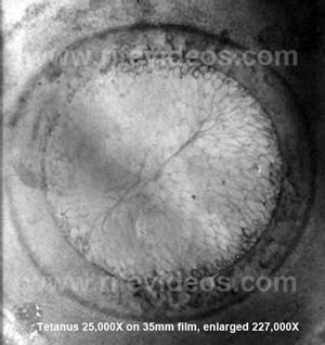
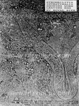
Under the universal microscope disease organisms such as those of tuberculosis, cancer, sarcoma, streptococcus, typhoid, staphylococcus, leprosy, hoof and mouth disease, and others may be observed to succumb when exposed to certain lethal frequencies, coordinated with the particular frequencies peculiar to each individual organism, and directed upon them by rays covering a wide range of waves. By means of a camera attachment and a motion-picture camera not built into the instrument, many "still" micrographs as well as hundreds of feet of motion-picture film bear witness to the complete life cycles of numerous organisms. It should be emphasized, perhaps, that invariably the same organisms refract the same colors. when stained by means of the monochromatic beam of illumination of the universal microscope, regardless of the media upon which they re grown. The virus of the Bacillus typhosus is always a turquoise blue, the Bacillus coli always mahogany colored, the Mycobacterium leprae always a ruby shade, the filter-passing form of virus of tuberculosis always an emerald green, the virus of cancer always a purplish red, and so on. Thus, with the aid of this microscope, it is possible to reveal the typhoid organism, for instance, in the blood of a suspected typhoid patient 4 and 5 days before a Widal is positive. When it is desired to observe the flagella of the typhoid-organism, Hg salts are used as the medium to see at a magnification of 10,000 diameters.
In the light of the amazing results obtainable with this universal microscope and its smaller brother scopes, there can be no doubt of the ability of these instruments to actually reveal any and all microorganisms according to their individual structure and chemical constituents.
With the aid of its new eyes - the new microscopes, all of which are continually being improved - science has at last penetrated beyond the boundary of accepted theory and into the world of the viruses with the result that we can look forward to discovering new treatments and methods of combating the deadly organisms - for science dose not rest.
To Dr. Karl K. Darrow, Dr. John A. Kolmer, Dr. William P. Lang, Dr. L. Marton, Dr. J. H. Renner, Dr. Royal R. Rife, Dr. Edward C. Rosenow, Dr. Arthur W. Yale, and Dr. V. K. Zworykin, we wish to express our appreciation for the help and information so kindly given us and to express our gratitude, also, for the interest shown in this effort of bringing to the attention of more of the medical profession the possibilities offered by the new microscopes.
REFERENCES.
ANDERSON T. F., AND STANLEY, W. M.: "A Study by Means of the Electron Microscope of the Reaction Between Tobacco Mosaic Virus and its Antiserum," Jour. Biol. Chem., 139: No. 1, 339-344, 1941.
ANDRADE, E. N. DA C.: "The Mechanism of Nature," G. Bell & Sons, Ltd., London, 1932.
APPERLY, F. L.: "Relation of Solar Radiation in North America," CancerResearch,I: 191-195, March '94'.
BATEMAN, 3. B., CALKINS, H. E., AND CHAMBERS, L. A.: "Optical Study of the Reaction Between Transferred Monolayers of Lancefield's 'M' Substance and Various Antisera,"
Jour. Immun.,41: 321-341, 1941.
BURTON, E. F.: "The Electron Microscope," Proc.Roy. Can. Insi., 5:Series 3A, Feb.10, 1940.
CORRINGTON, JULYAN D.: "Under the Microscope," Nat. Mag., 34:117-120, Feb.1941.
DARROW, KARL, K.: "The Renaissance of Physics," Macmillan Co., 1939.
DAVIS, WATSON: "Exploring A New World," Science News Letter,38: 227-230, Oct.12, 1940.
Same: Abr. with title, "30,000 Times Life-Size," Readers Digest, 37:13-16, Nov.1940.
DUNN, H. H.: "Movie New Eye of Microscope in War on Germs," Pop.Science,118: 27,June 1931.
ELDRIDGE, JOHN A.: "The Physical Basis of Things," 1934.
"Electron Microscope Magnifies Invisible World 25,000 X." Life, 8: 54, April 29,1940.
"Electron Microscope Shows 'Strep' Has Outer Membrane," Science News Letter,38: 301, Nov. 9,1940.
|