| Home | Accessory Kit | Marsh CD Collection | Library | Contact Us |
Dr. Rife's First Microscope |
 |
Introduction: This report was written by John Marsh back in 1966. John duplicated the report on a copy machine so the photos were of a poor nature. Where we could replace the poor quality photos with the original photos given in the report we have done so. Those photos which could not be replace have been left the way they were but we have tried to add some more photos that will help the reader to understand.
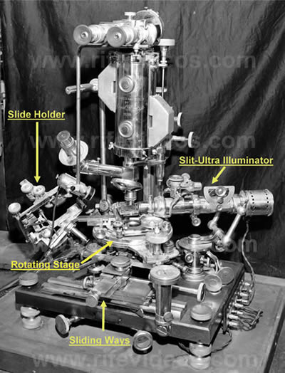
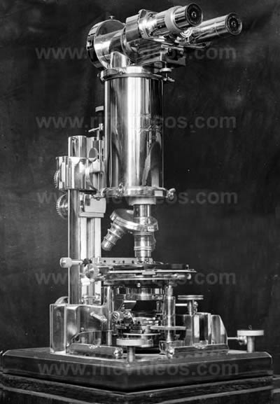
Since research to determine the cause and effect a cure of cancer is being carried out in various directions, it seems reasonable to explore every possible area toward a verified cause of cancer and hence, its proper method of treatment. Scientists today must re-examine the fact that a discovery was made of a virus of the dreaded cancer. That the discovery was made that such a virus was the cause of cancer. That the discovery was made that a method of treatment of electronics could electrocute the cause of diseases including cancer while the body cured itself. These discoveries of a virus causing cancer and treatment methods excited great interest among reputable men of the medical profession and professors and scientists and laboratory specialists as long ago as 1931. These facts have been founded on thousands of laboratory tests. The Rife Research Laboratory, in conjunction with doctors throughout the United States and Canada, worked to establish the techniques and methods to be used in controlled tests of the findings of Royal R. Rife's life study and devised methods of the cause and cure of cancer and other diseases. Royal R. Rife first conceived the idea of the Frequency Instrument in conjunction with his work in developing the virus microscopes. Over thirty thousand tissue slides were made and examined by Rife before he realized the futility of this effort. The virus was much smaller than the finest particles of the dye or colloidal stains and thus cannot be seen with standard laboratory methods. Since the diagnosis and determination of a cancer virus requires the high power of the Rife Virus Microscopes, a brief description of the details and principles of operation will follow on the Rife Universal. THE UNIVERSAL MICROSCOPE Born at the same time as the Electron Microscope, Rife's Universal was for his work only and has yet to be commercialized. Over a long period of 20 years, Rife designed and built in the Rife Research Laboratories five virus microscopes of power and resolution far beyond the laws of optical physics by using an entirely new optical system in which the rays do not cross as in the conventional scope. In the magnification of these virus instruments a variance from 10 to 60,000 times was seen. In contrast the commercial microscopes are inadequate for the observation of any of the filterable viruses of disease. The electron microscope kills them instantly and their forms change into dried up round balls and other distorted oblong shapes. Rife's virus microscopes enable the observer to study all viruses while they are living in their own true chemical colors which identify them as well. The cancer virus was found to be less than 1/20th of a micron long. Thus we see the need for such an invaluable instrument that would enable us to progress farther into this important field of endeavor. A description of the Universal, the most powerful of Rife's virus microscopes follows: The Universal Microscope, which is the largest and most powerful of all light. microscopes, was built and devised by Rife in the Rife Research Laboratory in 1933 which has advanced the art of microscopy to a state that has never been equaled yet. The Universal derives its name from its adaptability to all fields of microscopical work. It is fully equipped with three separate sub-stage light condenser units for monochromatic beam light, polarized light, and dark field light which may be selected and moved into position. An additional unit for slit-ultra illumination is mounted at the right side above the base. This has proved invaluable in dark field examinations. The patented Rife Microscope lamp with magnifier is positioned in the center of the base. Between the illuminators and the stage there is a retractable pair of counter-rotating wedge shaped prisms. These prisms are used to select a portion of the visible light spectrum which passes through the specimens being examined. When the light of the visible spectrum passes through these prisms, it is split into many beams. Many are refracted to the side while a portion of the beams are then allowed to pass through the prisms and go to the stage above and illuminate the specimens in their own translucent and or luminescent chemical constituents. The mounting arrangement permits each of the two prisms to be rotated in opposite directions by a vernier control throughout 360 degrees. This vernier adjustment measures the angle at which the variable angle of light incidence is applied to and from the source beam of illumination. Since only a portion of the spectrum is visible at any time, it is possible to select any portion from one end of the visible spectrum to the other. When that portion of the visible spectrum is reached where both the organism observed and the color band presented - vibrate in exact accord, a definite characteristic color is emitted by the observed organism at resonant light frequencies. The Universal Microscope is two microscopes in one unit where the lower or standard unit extends to the left side and allows the observation of normal study over the same system. When moved aside, the light progresses up and through the main body tube to the oculars. The original system contained 14 lenses and prisms. Three are in the high-intensity incandescent lamp, four in the Risley prism, and seven lenses are in the achromatic condenser.
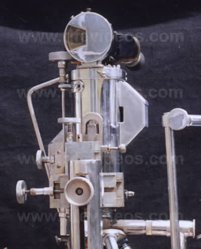
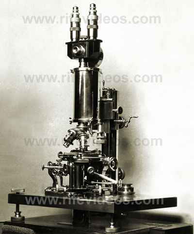
The Slit-Ultra Illuminator is shown at the right side and the Slide Holder for crystal angle measurements at the left side can be moved in five planes with compound dialed movements. The sliding ways mounted at the front of the base are used to control movements of the Lamp Assembly. The dialed movement is at two right angle planes with rotative movement also possible in the attaching nut fastener. The jeweled dial indicator to record the movement of the stage fine adjustment is shown at the right which works off of a taper attachment. VIEW OF LAMP ASSEMBLY AND REAR VIEW OF BODY
As Published by: The Smithsonian Institution in 1944 in the Annual Report of the Board of Regents and printed by the U.S. Government Printing Office, Washington, D.C. 1945 as their Publication Number 3776 written on pages 193 through 219 with 5 additional Plates entitled: "THE NEW MICROSCOPES" By Dr' R.E. Seidel, M.D., and M. Elizabeth Winter, both of Philadelphia, Pa. and the same name and same authors as published by: U.S. Government Printing Office in Publication Number 3781; and by Journal of the Franklin Institute, Vol. 237, No.2, February, 1944; Pages 103 thru 130. VIEW OF UNIVERSAL MICROSCOPE COUNTER ROTATING STAGE
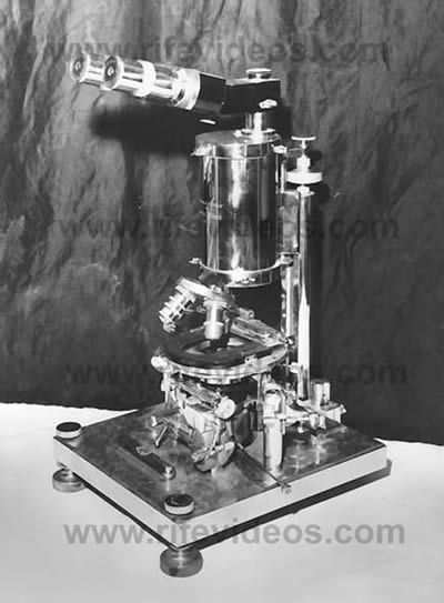
The magnifier is used to read the quarter minute arc divisions for precision location of the 360 degree continuous movement as desired. There is also a stop to lock the dual stage in any position desired. The dial type knurled knobs on the left in the photo [and on the right in the blueprint] are the reduction movement for the vertical fine adjustment of the stage which goes into a tapered reading on the dial indicator. The entire assembly can be removed with one knurled nut which is shown on the extreme right side and which is a separate part and is merely resting on the metal support. Another vernier is provided for the upper stage for precision settings of this stage also. The rays of light refracted by the specimen enter the objective lens and are carried up the tube in parallel waves through 21 light bends to the ocular eyepieces. A tolerance of less than one wave length of visible light is permitted in the core beam. In the standard optical microscope, the light rays tend to converge in the center of the body tube which causes an interference band of refraction thus limiting the power of magnification. In the Rife Virus Microscopes, as the light frequencies are about to cross each other, a specially designed quartz prism is inserted which serves to separate the rays to a parallel system. The prisms in the body tube are adjusted and held in alignment by micrometer screws in special tracks and are separated by a distance of about 30 millimeters. Thus the greatest distance that the image is projected either through quartz or air, is 30 millimeters instead of the usual 160 to 190 millimeters employed in the ordinary microscope. The new parallel ray principle used in the Universal Microscope provides the highest magnification and resolution known and serves to virtually eliminate all chromatic and spherical aberration. The stage is a double rotation device graduated through 360 degrees in quarter minute arc divisions. The upper segment carries the mechanical stage with a movement of 40 degrees, and the body assembly which holds and adjusts the slide sideways and in and out as desired. The main supporting stage is free to rotate by finger tip control through 360 degrees. The fine adjustment is operated by moving the stage up and down being 700 times more sensitive than ordinary microscopes and requires from 5 to 30 minutes to focus in. The time required is only a slight inconvenience compared to the many years of research and the actual results obtained in isolating and looking upon living disease causing virus and other micro-organisms in their true form.
The Number 5 virus microscope was built for an English doctor with quartz optics with a rotatable body tube similar to the Universal and with an up and down movement on the fine adjustment located below the stage.
The Number 4 virus microscope was built for Rife's own use and traveled extensively with Rife on trips. The coarse adjustment similar to that on the Universal Microscope is shown at the rear on top of the body tube support.
The Number 2 virus microscope also traveled and was used by Dr. Kendall in Chicago at the Northwestern University Medical School. It was an improvement over the No.1 scope.
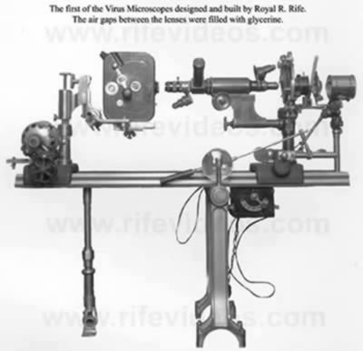
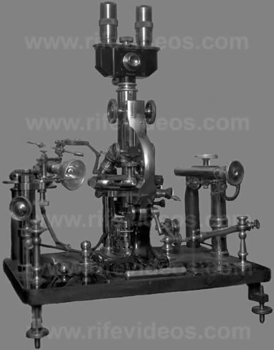
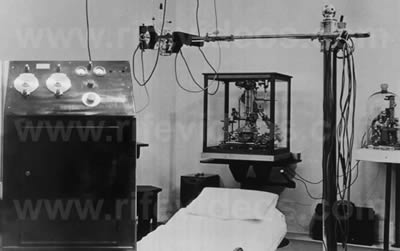
The first Rife virus microscope was published in a joint article by Dr. Kendall and Rife in California and Western Medicine, Vol. 35, No.6, pp 409-411 under a 1930 copyrighted photo in December 1931. A movie camera is shown at the left for micro-photography. The air gaps between the lenses were filled with glycerin. Rife's patented lamp is at far right.
Of great use in the slide preparation and analysis of frequency ranges was Rife's Petrographical Micropolariscope which he designed and had Lutz build. Here it is shown on his micro-manipulator which he built for microorganism study. Polarized top & below.
With the Rife Virus Microscopes a trained operator can determine whether a patient has cancer in a short time as well as other virus type diseases by their own characteristic chemical colors. It has also been found that in many types of epithelioma that the carcinoma tissue carries no conductivity. Thus with a pendulum galvanometer, the area can be outlined and the location of the tumor can be determined without the use of harmful deadly X- Rays or X-Ray Photographs. All pathogenic diseases can be eliminated with preventative treatments using the Frequency Instruments. Rife said, "We find, in 98% of carcinomatous individuals in the monocytes of their blood, granular forms. Malignancy is a blood disease and one stage of this granular form can be grown into carcin- oma by growth on Kendall's "K" Media made from pig gut and a tyrode solution. From a Crytomyces Pleomorpha I can transfer any of these stages back into a BX and produce a tumor from which a BX can be recovered."
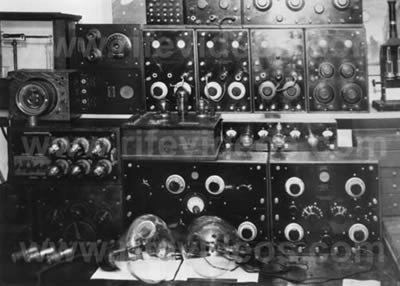
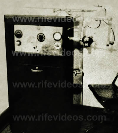
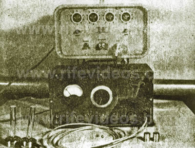
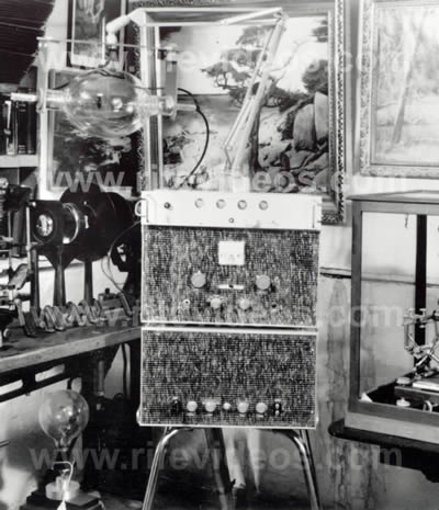
Royal R. Rife's discovery of the various electronic characteristics of organisms led him to believe that each kind of microorganism was electrical in nature and should have a resonant frequency determined for each individual type of chemical combination. His isolation of positive, -neutral, and negative bacteria showed that only combinations of + and - bacteria would reproduce. His first attempts to electronically destroy them consisted of unsuccessful tests with infrared and ultra-violet rays. Some local, surface destruction was noted but below the surface, penetration was impaired. He next tried Audio and Radio and frequencies. He thought that somewhere in the spectrum a harmonic or resonant frequency would resonate with the vibratory rates of disease organisms. He believed that if such a frequency could be found and applied, the rays would be fatal to the disease organisms. The first frequency instrument was designed and built in 1920. The instruments frequencies were transmitted and directed through a ray tube and was complicated to operate. Much of the research was also being carried on for the elimination of the virus of polio and tuberculosis as the work went into the 1930 period. Since the frequencies which would affect the virus were unknown, Rife proceeded with cultures on a trial and error basis under the virus microscopes. Rife and his assistants made test after test until at last initial success was found. Guinea pigs inoculated with the bacillus of tuberculosis and subjected to the frequency instrument at one particular frequency resulted in the organism being killed. At this time a problem even more difficult to solve became apparent. Even though the organism had been killed by the resonance of the proper frequency, in several cases the inoculated guinea pigs died of toxic poisoning. Three years were spent in determining the answer to this problem. A virus was suspected from the tuberculosis bacillus. Subsequent studies demonstrated that this was exactly the case. Rife used a 000 porcelain Berkefeld filter to isolate the virus in pure form unmixed with contaminating organisms by taking media from many plated cultures of the bacillus of tuberculosis. That Rife managed to find the proper frequencies for both forms of T.B. and Cancer and other virus diseases as well as devising techniques working with Dr. Arthur I. Kendall of Northwestern University is a great tribute to his remarkable versatility. "We have been able to devitalize the virus and bacillus of tuberculosis with radio waves since 1930," Rife said, "but we could not use the treatment until we had done more work to make it possible for man's use. After being subjected to the waves, the organisms will not grow in culture flasks and will cause no disease when injected into guinea pigs." With transmitted RF frequencies through a large rare gas filled ray tube, the destruction of the organisms by rays and frequencies is described as being similar to the phenomenon of a combination of transmitted electronic energies and the coordinative resonance of resonant fundamental frequencies or their harmonics. This is likened to fragile glass which is shattered by a sustained note of music tuned to a resonant pitch or where a potential energy charge is built up to exceed that level of energy which the micro-organism can cope with and still live. The following photo shows the many dials and controls used in the original Rife Frequency Instrument with two Ray tubes of the early type where Coolidge type electrodes were used to direct the electronic energies:
RIFE RAY TUBE FREQUENCY INSTRUMENT
Above is the early Rife Frequency Instrument used in the clinical work of a Special Medical Research Committee of the University of Southern Calif. 1934. Rife often demonstrated "that his Frequency Instrument had the power to kill germs that cause B-Coli, E-Coli, Tuberculosis, Cancer and other infectious diseases and human disorders, without harm to human tissues. Working with Dr. Walker of the Hooper Foundation in San Francisco, Rife isolated the virus of leprosy and also devitalized it. This effort was arranged by Dr. Milbank Johnson, M.D. and. Dr. Karl Meyer, M.D. Director of the Hooper Foundation of the University of Calif. Rife was also aided in pathology by Dr. Foord, M.D. through the requests of Dr. Milbank Johnson, M.D., Pig studies were made by Dr. Foord in 1933 for Royal R. Rife. At this time Dr. E. C. Rosenow, M.D. of the Mayo Clinic aided Rife and Kendall in further collaboration. Dr. Karl Meyer, M.D. was a member of the Special Medical Research Committee of U.S.C. Development of the Rife Ray to the point where it can be used on human beings was a proven fact back in 1934. Tubercular and Cancer patients treated in private practice have recovered. Most cases respond within a period of one to two months and the diseases are instantly rendered non-infectious.
The isolation of the cancer virus was an accomplishment in which Royal R. Rife took a great deal of pride. In 1931 he discovered the transformation of the cancer virus and the successful treatment for cancer virus by his actual observation under the Universal Microscope while applying the frequency instrument and its lethal electrocuting energy. The major portion of the cancer tests of the tumors used initially were from the Paradise Valley Sanitarium in National City, California. The pathology was checked through their laboratory as malignant. Dr. Hamer, M.D. worked with Rife and Ben Cullen (in 1937-38). The methods and procedures follow: An un-ulcerated breast mass checked and passed as having cancer malignancy by the Paradise Valley Sanitarium and the Rife Research Laboratory in Point Loma was placed in a triple sterilized container. Rife next took a small portion from the un-ulcerated breast mass and sealed it in a test tube with "K" media applying a two inch water vacuum. It was then ionized for 24 hours in an argon gas filled loop. Upon examination of the solution in the test tube, it was found to be teeming with "BX" or cancer virus, which were the most highly motile and the smallest in size of any of the viruses previously isolated. When examined under the Rife Virus Micro- scopes after passing through 000 porcelain Berkefeld filters, all cancer virus refract a purplish-red color with the monochromatic beam which is a positive diagnosis in all cases. The "BY" virus was identified by Rife as a larger form of the Sarcoma group. The method of inoculation of experimental animals with "BX", the virus of cancer is as follows: The animal is first shaved and sterilized with alcohol and iodine solution at the point of inoculation and placed under partial anesthesia. This avoids subjecting the animal to shock. An extra long, very small needle was used. The needle was filled with sterile petroleum jelly and a hypodermic was then filled with the inoculum of the “BX” and the needle placed on the syringe. The needle was inserted no less than 30 millimeters from the point of inoculation under the epidermis of the albino rat. The point of inoculation in most cases was the mammary gland of the experimental animal. Often these growths or tumors exceeded the weight of the animal. Rife surgically removed the tumor, treated the animal with the Rife Ray and the animal recovered. The "BX" was again recovered and injected into another animal in tests to further prove the cause of cancer. An important factor and check of the pathology is to make at least 10 transplants from the initial isolation into the "BX" media. These transplants are made at 24 hour intervals into the original "K" media. With ionization this increases the virulence and speeds the growth of the tumors. With these experiments that have been repeated on over 100 experimental animals, we are convinced that this procedure definitely proves the cause of malignant carcinoma commonly referred to as cancer; the virulence and the pathology of a virus isolated from medically diagnosed “BX.” If there are any workers interested in following this technique, we will furnish them with all of the basic principles involved. However, it is beyond the scope of the average microscope to visualize these minute "BX" cancer viruses.
The actual cure of cancer in experimental animals and humans occurs with the use of our Frequency Instruments. To attain these astounding results, a long and tedious process is started to determine the precise setting of the Frequency Instrument which was originally termed M.O.R. or the Mortal Oscillatory Rate of the virus of BX. When the setting is found, it is repeated 10 consecutive times after the Frequency Instrument has been dialed back to the same reading before a specific M.O.R. or frequency is recorded as seen under Rife's Virus Microscopes.
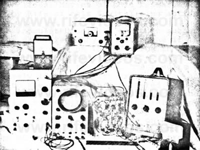
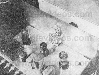
The test instrument at the right with the vertical dials is a Hewlett-Packard Frequency Counter for the precise calibration of audio frequencies. This instrument is accurate to one beat in 100,000 cycles per second having a special built in time base. The VTVM at top left is another Hewlett-Packard instrument noted for its high quality. Other instruments used for testing are an Oscilloscope to observe the vacuum tube output waveform, a multimeter and a db VTVM. When the mortal oscillatory rate or potential energy level is lethally reached, the BX form of the virus appears to blow up and disintegrate in the field and form, at times, a group of agglutinophilic clumped masses or electrostatic lines. Movies of this research were made by Rife of this devitalizing action. The inoculated animals are then subjected to the same frequency to determine if the effect is the same on the BX virus in the tissues of the experimental animals. It was always found to be the same. The results were precisely identical with experimental animals as with the pure culture filtered virus BX slides. These successful tests were conducted over 400 times with the albino rats experimentally with controls before any attempt was made to use these frequencies on human cases of carcinoma, sarcoma, leukemia, and other forms of neoplastic and parasitic trouble. Rife's first human clinical work on cancer was completed under the direction of Dr. Milbank Johnson, M.D. Chairman of the Special Medical Research Committee of the University of Southern California which included Dr. George Dock, Professor of the Theory and Practice of Medicine, Medical Dept. Tulane University of Louisiana, New Orleans; Dr. Charles Fischer, M.D. of the Children's Hospital in New York; Dr. Wayland Morrison, Chief Surgeon of the Santa Fe Railway, M.D.; Dr. Karl Meyer, M.D., Director of the George Williams Hooper Foundation, Dr. Arthur I. Kendall, Professor of Bacteriology at Northwestern University Medical School in Chicago. The research was conducted at the Scripps Home which was rented over the summer of 1934 for cancer & T.B. cases. 14 out of 16 were cured in 70 days Rife said. The work was also confirmed by Dr. Couche, M.D., and others verifying and checking the work. Dr Kendall reported that Dr. Alvin G. Foord, M.D., diagnosed Tom Knight, one of the cancer cases and his letter to Rife states: " Tom was an excellent case because the moulages before and after were so striking; there were the actual models of the lesion in the first days and after some 75 days: also the biopsy report as I understand it was beautiful in that Dr. Foord, so I understood, diagnosed the lesion in the first days and after some 75 days as a malignant epithelioma.." and in a letter to Mrs. Bridges: "I have written to Dr. Johnson telling him of the one case I can talk about intelligently: Tom Knight. Roy (Dr. Rife) will tell you about Tom: he seems to me to be the most important case of the entire series because his tumor was on the cheek, where it could be seen, watched and measured from the start to the finish. This I have done reciting the actual measurements, and details of treatment and of pathological examination." The treatments consisted of three minutes duration which was set on the Mortal Oscillatory Rate with transmitted Radio Frequencies coming from a large Rife Ray tube antenna. Destruction of the organisms by the rays and frequencies is described as being similar to the phenomenon of a combination of transmitted potential electronic energy and the coordinative resonance of critical frequencies. This is electromagnetic electrocution where a potential energy level is reached that is lethal to the cell walls of the microorganism but still harmless to the human cell wall which is more resistant to this critical energy level and survives. The treatment time interval was divided into two groups; half of the patients were treated every day; half of the patients were treated every three days. Those treated every three days recovered faster than those treated daily because the lymphatic system was not overloaded. It had a better opportunity to absorb and cast off the dead particles of the BX or cancer virus and the dead particles of the Bacillus and virus of Tuberculosis. No rise of body temperature was perceptible in any of these cases above normal during or after the Frequency Instrument treatments. No special diets were used in any of this clinical work, but we think a proper diet compiled for the individual would be of great benefit where the food used would have been grown on properly prepared and mixed soil with inorganic and organically used chemicals. Food grown from depleted soils can make us sick just as food grown from soils that are chemically unbalanced or where one or several elements are in excess.
Other doctors and important men observing results were: Dr. James B. Couche, M.D., Dr. Ray Lounsberry, M.D., Dr. E. F. F. Copp, M.D., Dr. Thomas Burger, M.D., Dr. Arthur I. Kendall, PhD, Dr. Joseph Heitger, M.D., of St. Louis, Mo., Dr. O. C. Grunner., M.D. Pathologist and Head of the Archilbald Cancer Research Committee of McGill University of Montreal, Canada, Dr. E. C. Rosenow M.D., Head of Bacteriological Research at Mayo Clinic, Rochester, Minn., Dr. S. Fosdick Jones, Dr. C. M. Hyland, Dr. V. L. Andrews, Dr. Alvin G. Foord, Dr. F. C. E. Mattison, Dr. E. M. Hall, Dr. C. W. Bonynge, Dr. E. W. Butt, Dr. A. S. Hoyt, Dr. R. W. Lamson, Dr. A. H. Zeiler, Dr. R. W. Hammack, Dr. G. D. Maner, Dr. Ellis Jones, Dr. O. O. Witherbee, Dr. Harold Witherbee, Dr. B. O. Raulston, Dr. Linford Lee, Mr. Albert Ruddock, Mr. Richard Winter, Dr. George Kress, Mr. Aubrey Davidson, Dr. W. H. Scoins, Dr. Walter V. Brem, Dr. Allen B. Kanavel, Dr. Samuel J. Mattison, Mr. J. Brandon Bruner, Miss Kendall and Miss Couche, Henry Seiner, Ben Cullen, Jack Free, Philip Hoyland, Verne Thompson and many others. With the passing of Dr. Milbank Johnson, M. D. and Mrs. Bridges, two of Rife's closest friends, and with the organization of the Beam Ray Corporation for the purpose of marketing Rife's Frequency Instruments which had its winning in a long Court fight but also its lost finances to sustain such a struggle, the officers decided not to sue and gave up against the power of the A.M.A. where the doctors were forced to give up the Frequency Instruments they had received from Beam Ray Corporation. of San Diego. Dr. Couche and Dr. Arthur Yale, M.D., continued to use the Frequency Instruments and cured countless cases until they both passed on in about 1959. Other doctors also used the Tube Type Frequency Instruments. Other clinics were held in 1935 through 1937 by Dr. Milbank Johnson, M.D., using Rife's Frequency Instruments. Great success was attained. In 1950 John Crane heard the story and decided that something should be done about it as he felt that too many people were dying needlessly. Crane hired Verne Thompson to build six Rife Frequency Instruments with a wiring diagram he secured from Dr. O. C. Gruner of Canada.
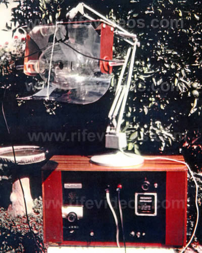
The reason that six were built was to qualify them with the Federal Communications Commission who also tests them for six months prior to their release for stray radiation of radio frequencies. At this time Dr. Charles F. Tully, D. D. S. decided to use one of Dr. Couche's Frequency Instruments and bought it from him and made history by curing mouth diseases and strep and staph infections as well as other sicknesses. Rife gave him his quartz window ray tube as shown:
Dr. R. E. Seidel, M.D. wrote, "Under the Universal Microscope disease organisms such as those of tuberculosis, cancer, sarcoma, streptococcus, typhoid, staphylococcus, leprosy, hoof and mouth disease, and others may be observed to succumb when exposed to certain lethal frequencies peculiar to each individual organism." Crane worked closely with Dr. Couche and watched some of the cancer cases clean up. One was Mrs. James Russell Jones and another was Mrs. John E. Marsh. Both well after five years as of 1954 approximately. Crane next tried to get permission from the Public Health Dept. to market the instruments and ran into a stone wall. The A.M.A. controlled puppets would have no part of it. Their own experts said it was safe to use but the Public Health Board said it was unsafe and denied the application. Crane was not discouraged and then set about to simplify and cut costs on a production model. It was reasoned that the transmitter portion and tube could be eliminated and in place substitute a transducer direct to the body. After several experiments in which some flesh had been burned, the small type "tube-less" Frequency Instrument was born. Only the audio oscillator portion now remained which was calibrated with a Frequency Counter. Two firm names were set up: Allied Industries and Life Lab Inc. by Crane. [We would like to point out that Dr. Rife did not approve of these new "tube-less" frequency instruments. Where John Crane and John Marsh went wrong is that they quit using the RF carrier frequency used in the original Beam Ray Clinical instrument. Dr. Rife really had no problems using a metal antenna (metal hand disks or cylinders and footplates) but the removal of the RF carrier is what really upset him. Dr. Rife really didn't know how Philip Hoyland's instrument worked but one thing he did know is that it had to have the RF carrier frequency to work properly and John Crane removed it. Below is Bertrand Comparet’s, Dr. Rife's attorneys, statement in regards to these new instruments: COMPARET: “And I asked Rife, because I thought Rife would certainly say that the way Crane was working on it then was still using the Rife principle, but he indignantly denied it.” Dr. Rife knew that his frequencies which devitalize microorganisms were not audio frequencies. Yet, John Crane and John Marsh were taking the audio frequencies from Philip Hoyland's machine, ignoring the RF carrier frequency, and telling everyone that the audio frequencies were Dr. Rife's original M.O.R.s. It is understandable, in view of this, why Dr. Rife was upset and blew up. Nevertheless even though these new small instruments only used audio frequencies they did help a lot of people with many ailments as John Marsh describes in his report. Today many people use these low audio frequencies with very good results but this does not change the fact that these low audio frequencies are not Dr. Rife’s true M.O.R. frequencies.] The original audio oscillator was a Hartley but others were available and all produced the desired frequencies. And so many were tried out experimentally on cultures of E-Coli under the Rife Microscopes. [When Dr. Rife was asked in his 1961 deposition about this small pad instrument that Crane and Marsh built he made this statement: Question: Have you ever made or observed a test of the effect of the electronic frequency-generator, of the type produced by John F. Crane, one of the defendants in this case? If so, tell us the kind of test or tests, who made such test or tests, and what result you observed. On the first of January 1960 Crane set up Rife Virus Microscope Institute for the purpose of advancing the state of the art of the tube type and small type Frequency Instruments as well as the virus microscopes and related research. Consulting Electronic Engineers were hired and worked to develop the necessary circuits. A glass blower was hired to fashion the newer and bigger Rife Ray tubes which were to be powered by more powerful Rife Frequency Instrument transmitters with the Tektronix Square Wave Generator. Medical and Chiropractic doctors were contacted to conduct research with the Frequency Instruments and approximately 70 small type frequency instruments were produced for doctors and people where the "Training of how to operate the Frequency Instruments" was sold and the device was loaned for research evaluation only and Lab operating manuals were furnished along with a training program to acquaint those with the techniques required to attain successful results. No claims were made as stated in the signed contracts.
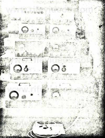
The results started pouring in. Many were getting benefits. Diseases which they had for years suddenly started clearing up. The new Frequency Instruments were straightening out their weakened cell structure while their bodies gradually eliminated the parasitic trouble. Frequency Instruments were taken by John Marsh to Dr. Robert P. Stafford, M. D., in Ohio and to Dr. Leonard R. Chapman. M. D., D. O., D.C., in Vista, California and to Dr. Edward M. Jeppson, M. D., in Utah. Agreements were made for their research evaluation. Dr Eason, N. D. of Utah also received a Frequency Instrument:
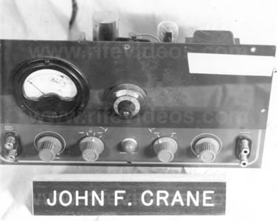
This device was seized and given to the Food and Drug branch of the Dept. of Health, Education and Welfare. The Eason Frequency Instrument had direct reading dials in that when the audio oscillator was tuned the frequencies produced by it were read directly on the four dials above which made the instrument self calibrating and in correct precise alignment at all times.
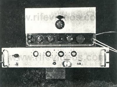
Dr. Chapman's instrument had an eleven position switch at the top center with a band tuning dialer at top center above which was Model SQ-2-10; four of these units were made. The circuit could not be designed for less than $1000.00. The Frequency Counter shown below it was not included. It is a commercial Westport Co. Frequency Counter costing $315.00. Dr. Chapman, M. D., was a member of the Rife Virus Microscope Institute. Dr. Ruth Drown, D.C., a member of the Rife Virus Microscope Institute and she used the frequency instrument. She was widely published after a lifetime of work adding a great contribution to science with her photo-cell electronic developments and photos. Dr. Charles Bunner, D. C., a member of the Rife Virus Microscope Institute also used the frequency instrument. Dr. R. C. Colburn, D.C., a member of the Rife Virus Microscope Institute used the frequency instrument. Dr Colburn was planning on giving up his practice because of the state interference with his business from use of the frequency instrument. And then we have the John Doe Doctors and People who say - yes I want the Frequency Instrument and they work wonderfully well but please don't mention that I have it as I can't afford to lose my job, I can't afford to suffer the losses in my business, I am afraid of the American Medical Association and the Drug Monopoly (and they use the Frequency Instruments, get well, and are kept confidential.). Dr. Adolphus Hohensee, M.D., D.C, N.D., a member of the Rife Virus Microscope Institute was also an ordained minister of the gospel. The copyrighted version of the Frequency Instrument is a modified commercial unit. It has performed with amazing success curing case after case and is shown below:
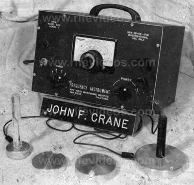
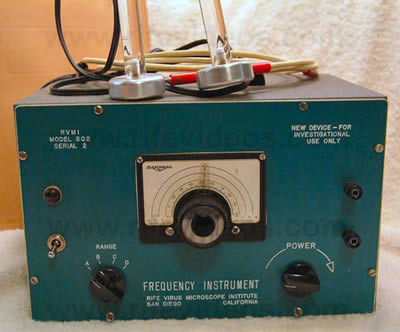
The words on the upper left [in the photo of the first instrument] read: "RVMI Model SQ-2" "Serial 1" and on the upper right: "New Device - For Investigational Use Only" The RANGE switch is at the lower left while the Power switch is at the lower right. The range of sound energy and potential energy level transducers are shown in the various sizes produced at the front in front of the device. This instrument weighs 12 lbs. without the transducers so that it may be readily moved about without difficulty. Green engraved plastic front was used in this series of the experimental tests. VIEW OF RESEARCH FREQUENCY INSTRUMENT
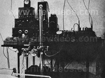
The Rife Virus Microscope Institute was in the development and research testing of the tube type new instrument shown above with the Tektronix Square Generator missing from the assembly. This work was being conducted in 1960 where tests on mice with cancer were received from the Institute for Cancer Research in Philadelphia. The frequency counter was located in the center in later configurations with the transmitter on top. Special ray tubes mounted on flexible arms transmitted the energy of the Frequency Instrument. The remainder of this report, not included, dealt with all the various legal appeals that both John Crane and John Marsh made in regards to their 1962 Court Trial. DATED: June 10, 1966 |