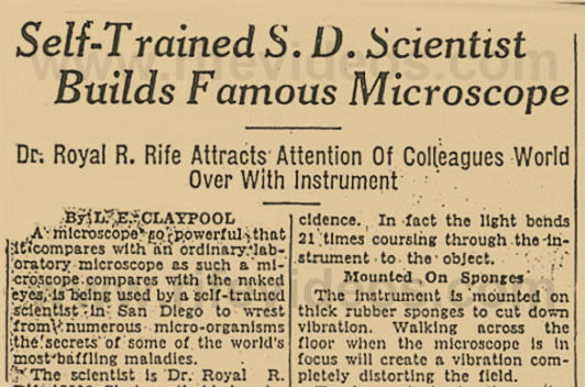
Self-Trained S.D. Scientist Builds Famous Microscope |
Self-Trained S.D. Scientist
Builds Famous Microscope
Dr. Royal R. Rife Attracts Attention Of
Colleagues World Over With Instrument
California Newspaper, April 16, 1934
By L. E. Claypool
A microscope so powerful that it compares with an ordinary laboratory microscope as such a microscope compares with the naked eyes, is being used by a self-trained scientist in San Diego to wrest from numerous micro-organisms the secrets of some of the world's most baffling maladies.
The scientist is Dr. Royal R. Rife, 2500 Chatsworth blvd., who recently built the microscope and thereby attracted the attention of scientists all over the world.
NO FOCUS CHANGE NEEDED
The microscope has a power range of 5000 to 20,000 magnifications. The instrument has 5,087 parts, weighs 200 pounds and has attachments making it possible to use every phase of microscopy in viewing an object without changing focus.
Because of its great power the Rife microscope used solely for the study of filter-passing organisms. They are called filter-passing because they are so small they will pass through the finest porcelain filters.
But it is not magnification alone that enables one to see these minute bodies. A special source of illumination is necessary without which the organisms could not be seen no matter how great the magnification.
LIGHT ANGLES "BENT"
Variable beams of monochromatic light are bent at an angle of incidence corresponding to and coordinated with the vibratory rate of the organisms being examined. In this manner the minute granule, which is too small to absorb ordinary stain, is "stained" in its true chemical color. This color is always the same under the proper conditions and thus serves as a definite method of immediately classifying the filter passers.
The apparatus used in this illumination system consists of four rotating quartz prisms, mounted beneath the substage condenser. Proper manipulation of these prisms bends the monochromatic beam at the desired angle of incidence. In fact the light bends 21 times coursing through the instrument to the object.
TO MOUNTED ON SPONGES
The instrument is mounted on thick rubber sponges to cut down vibration. Walking across the floor when the microscope is in focus will create a vibration completely distorting the field.
To demonstrate to the writer the microscopes powers, Dr. Rife placed upon the stage a fragment of a liver capillary. Under an ordinary microscope several of such capillaries may be seen. Under the Rife instrument one capillary is too large to be seen as a whole.
Dr. Rife explained that there are 5,000,000 red blood cells in a cubic millimeter of blood. Many of these can be seen in the field of an ordinary microscope. A single blood cell occupies the entire field of the Rife microscope.
Explaining it in technical language, Dr. Rife said: "The microscope embodies the transmitted light illumination, monochromatic beams of light, the dark field, slit illumination, and takes in the complete work of the polarizing microscope. Three illumination units are mounted on a track beneath the substage condenser. One is for dark field; another for polarized light and the third is for monochromatic and transmitted light.
"To use any of the units the microscopist simply slides into position the particular unit of illumination desired. The focus remains untouched."
With the aid of the microscope, which it took four months to build, Dr. Rife hopes to open new fields of research into pathogenic phenomena. Among the filter passing viruses the scientist has been studying are those of psittacosis and sleeping sickness. These he declines to discuss until he has submitted reports formally in the scientific magazines.
AT IT 30 YEARS
Dr. Rife is been engaged in scientific research for 30 years. Among the instruments he has developed is one known as the mechanical finger. It is an instrument which handles a quartered hair of a baby to lift into place minute objects for microscopic inspection. He also has a large collection of rats and guinea pigs for research. Into these he grafts malignant tumors and observes their growth and then operates an anaesthetically to remove them.
He also has what laymen sometimes call a death-ray machine (Rife Ray or Rife Machine). It is in the nature of a mechanico-electric bacteriophage. It is supposed to destroy micro-organisms by impact at their own vibrations. Much of Dr. Rife's work is done in collaboration with Dr. Arthur Isaac Kendall of Northwestern University, shown with Rife in the accompanying photograph. |

